

All premium Magento themes
at magentothemesworld.com!
Foundations of Medical Ultrasound Imaging
Ultrasounds in some drawings :
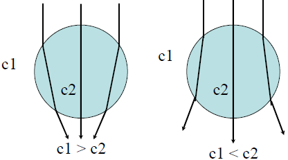
Refraction
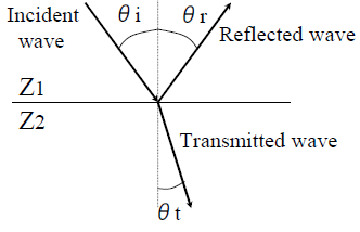
Reflection and transmission
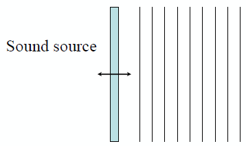
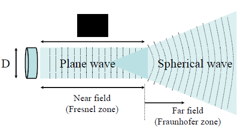
Plane wave
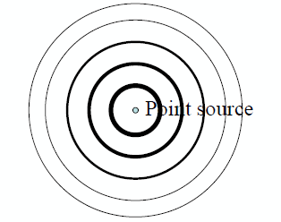
Spherical wave
In this page, we can try to explain the foundations of medical imaging by ultrasounds. For that, we will explain at first what is an ultrasound signal to after understand the basics principle, how we can make an image with those ultrasounds. To finish, we will see different possibilities offered by this method.
So, let's begin by this question :
What is
Ultrasound ?
- Frequency of ultrasounds are higher than the upper limit of hearing which is about 20kHz but this definition is not very clear, the frequency range stay very huge.
Now, what about physics of ultrasounds in the body ?
- Velocity of propagation :
- About 1540[m/s] in human body
- Each tissue has its own velocity.
- Way of propagation :
- Two way for the wave to propage : There are sperical and plane waves.
- In practice, the ultrasound is at first, a plane wave and it becomes a spherical wave, like it is explained in this schema.
In practice
- Wavelength : About 0.437[mm] in the body (3.5MHz)
Basic principle of ultrasound echo imaging :
This plan can resume this principle :
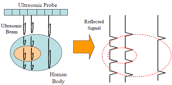
This plan can resume this principle :
Ultrasonic
waves are emitted by the ultrasonic probe. They propagated in body
tissues and each time the environment change, a part of wave is
reflected. The probe can receive those echoes. And by analyzing these
echos, we can make an image of propagation environment of ultrasonic
wave.
Many methods permit to improve how we make the waves to propagate. To obtain an image, we need ultrasonic beam. But, in human body, ultrasonic waves are very spread and they propagate as spherical waves. So we need to focusing waves. There are two main method for this. We can by example focusing wave energy in one point (the focal point) by using acoustics lens or "electronic lens", more precisely, electronic focusing. We can note that electronic focusing can be used in elission or in recepetion. These two methods matched to the two schemes following :
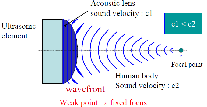
Many methods permit to improve how we make the waves to propagate. To obtain an image, we need ultrasonic beam. But, in human body, ultrasonic waves are very spread and they propagate as spherical waves. So we need to focusing waves. There are two main method for this. We can by example focusing wave energy in one point (the focal point) by using acoustics lens or "electronic lens", more precisely, electronic focusing. We can note that electronic focusing can be used in elission or in recepetion. These two methods matched to the two schemes following :
Acoustis Lens
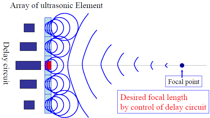
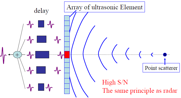
"Electronic Lens" respectively in emission and in reception
Now, take a quick look to use of this method.
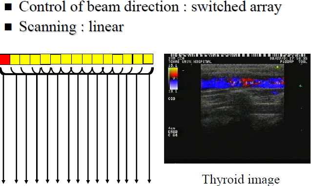
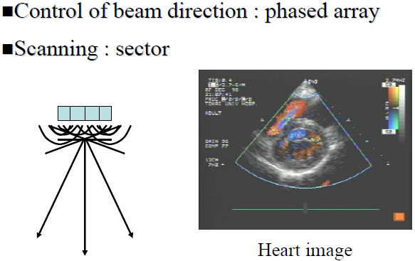
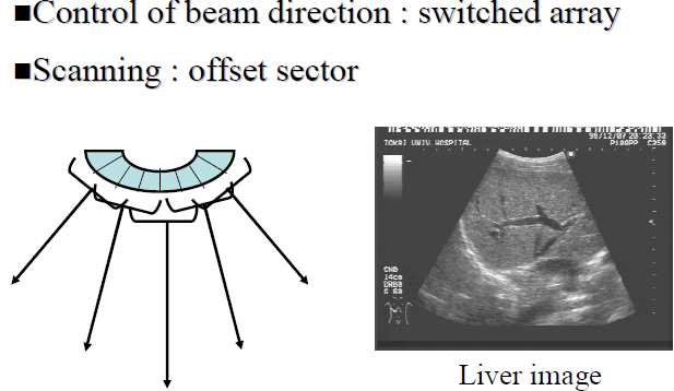
Different scanning techniques :
Several scanning techniques exist. By changing the probe form or use different control beam for the wave, we can observe different things. Just see examples below :
 |  |
 |
We can summarize those methods with table following :
| Element Array | linear | convex | linear | annular |
| Control of beam direction | Switched Array method | Phased Array method | Mechanical | |
| Scan | linear | Offset sector | sector | |
| Probe form | linear | Convex | sector | |
| Region of image | thyroid; breast | Abdominal region | sector | |